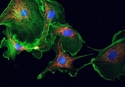
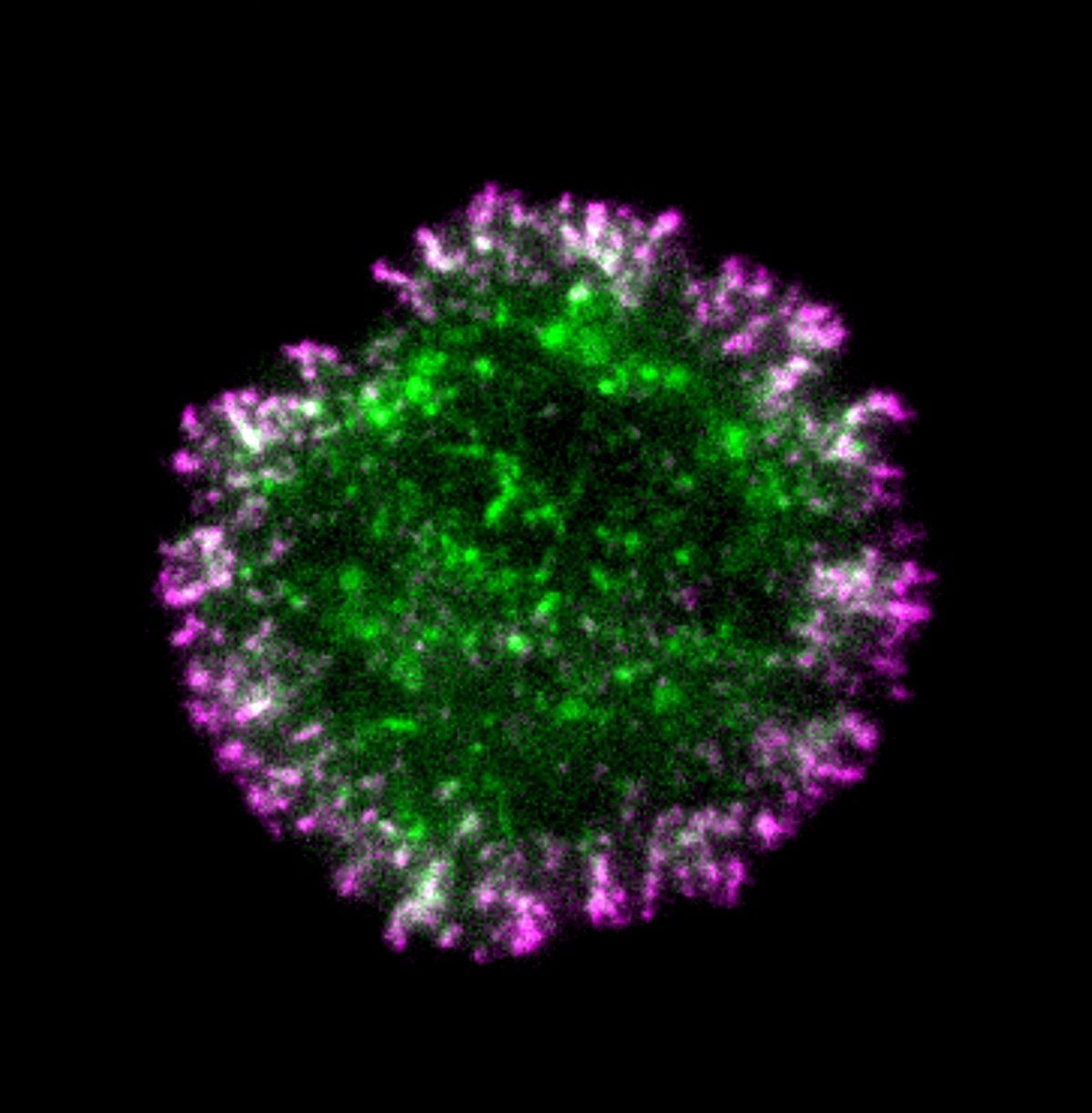
ABOVE: The cytoskeleton, in green, supports cell structure and promotes migration. © iStock, defun
Cells migrate during development, immune defense, and tumorigenesis, and this requires forming and dismantling attachments with the extracellular matrix and other cells. Force-sensing and adaptor proteins work together through a series of signaling cascades to activate surface molecules called integrins, which form adhesions at the cell surface that ultimately connect with extracellular targets. Most models of this process indicate that force-sensing proteins initiate these integrin-activating cascades, but early adhesion events, including whether the small forces present are sufficient to activate these proteins, are not well understood.1-3
In a new study published in eLife, researchers reported that the adaptor protein Cas promoted the initiation of cellular adhesion, suggesting a force-independent activation.4 Once activated through phosphorylation, Cas forms complexes with other signaling proteins that contribute to cell adhesion, migration, and proliferation. However, until now, scientists thought that these events occurred after the activation of integrin through mechanical sensation. “This really puts all the phosphorylation and sort of signaling events in the driver’s seat of the assembly of the adhesion,” said Jonathan Cooper, a cell biologist at the Fred Hutchinson Cancer Center and coauthor of the study. Understanding these early steps can help explain how cells recognize and form attachments in their environments and how cancer exploits this process to evade destruction.
See also “How Soft or Stiff Substrates Direct Stem Cell Differentiation”
“Focus has been on how cells become mechanosensitive, how cell adhesion became mechanosensitive through the recruitment of mechanosensitive proteins like talin, vinculin, and also other proteins,” said Sangyoon Han, a mechanobiologist at Michigan Technological University who was not involved in the study. “But not many studies were around to tell if there is any kind of premechanosensitive processes that act as a seed for the original binding of this mechanosensitive binding.”
Cooper’s team imaged adhesion formation at the cell surface interface using total internal reflection (TIRF) microscopy and observed that activated Cas clustered at this site almost a full minute before mechanosensing proteins arrived or β1 integrin was activated. They depleted Cas with siRNA and inactivated it to determine this protein’s role in the developing adhesion site. “When we remove Cas from cells, or inhibit its phosphorylation, or inhibit stuff downstream of Cas, the integrin clustering doesn’t really happen,” Cooper said. The Cas-depleted cells attached poorly to surfaces and were immobile.
Finally, the group identified a positive feedback loop between Cas and Rac1, which drives actin polymerization and is important in forming focal complexes that promoted cell adhesion formation.5,6 Cas phosphorylation activated Rac1, which in turn generated reactive oxygen species, which promoted additional Cas activation. Cas degradation regulated this loop.
See also “The Role of Adhesion Receptors in Cancer Cannibalism"
"It really nicely revealed all the sequence and dependence of very well known biochemical proteins in the focal adhesions, and they act as a seed for the oldest mechanosensitive protein recruitment and assembly,” Han said.
Han noted that while there were many unknowns left, including how Cas arrives at the cell edge, the study provides an important foundation for further research. “Now people can change their perspective on the focal adhesion assembly,” Han said.
References
- Wegener KL, et al. Structural basis of integrin activation by talin. Cell. 2007; 128(1):171-82
- Kim C, et al. Talin activates integrins by altering the topology of the β transmembrane domain. J Cell Bio. 2012; 197(5):605-11.
- Franz F, et al. Allosteric activation of vinculin by talin. Nat Commun. 2023; 14(1):4311.
- Kumar S, et al. Cas phosphorylation regulates focal adhesion assembly. eLife. 2023;12e90234
- Pasapera AM, et al. Rac1-dependent phosphorylation and focal adhesion recruitment of myosin IIA regulates migration and mechanosensing. Curr Biol. 2015; 25(2):175-186.
- Grobe H, et al. A Rac1-FML2 signaling module affects cell-cell contact formation independent of Cdc42 and membrane protrusions. Plos One. 2018; 13(3):e0194716.

